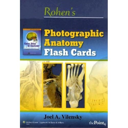
This set of 220 flash cards were developed with the primary aim of helping health profession students prepare for a practical exam in a typical medical gross anatomy and/or neuroanatomy course. Although there are many anatomy flash card sets available, none are based on photographs of cadaver dissections. The advantage of using photographic images rather than drawings for preparing for a practical exam seems self-evident.
All the photographs in this flash card set were derived from the sixth edition of Color Atlas of Anatomy: A Photographic Study of the Human Body by Johannes W. Rohen, Chihiro Yokochi, and Elke Lütjen-Drecoll (2006). The photographs in that atlas are exquisite in their detail.
 Before trying to identify a structure, the student should first become oriented—that is, determine what is medial, lateral, superior, inferior, anterior, and posterior. The student should use the same approach when taking the exam itself. These cards lack the three-dimensional definition that cadavers offer; thus, identifying structures on these cards is probably harder than identifying the identical structures on a cadaver. However, the dissections used for these photographs are so good that this liability is not significant and is equally prevalent on flash cards based on drawings.
 The front of each card typically has three or four structures to identify. The correct answers are identified on the back of the card. Further, each answer includes some important anatomical characteristic of that structure that may help the student identify it more readily or answer a written question about it. Also on the back of the card is the page number of the atlas that contains the image shown on the front. Thus, the student can easily obtain more information about each image.
Performing well on a practical exam is a skill that some students find difficult to master. Students will find these cards helpful in acquiring that skill.